Musculoskeletal Imaging
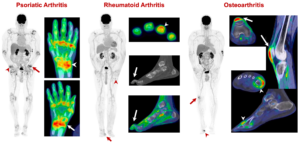
Total-Body PET/CT in Arthritis
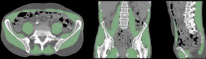
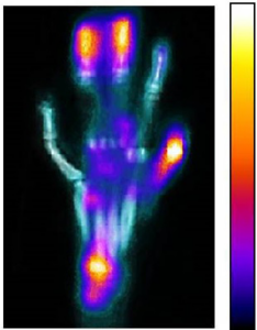
Our group is developing novel imaging methods to assess the systemic and local burden of Rheumatoid Arthritis, Psoriatic Arthritis and Osteoarthritis. We performed the first-in-human studies using a total-body PET/CT scanner in the arthritic population. These scans used an ultra-low-dose protocol, and enabled the visualization of the inflammatory activity associated with arthritis systemically. We believe that these promising results will lead to an improved understanding of arthritis and autoimmunity, and contribute to a new paradigm of using systemic molecular imaging methods to monitor disease activity and treatment response.
Reference: Y. Abdelhafez, S. Sarkar, H. Hunt, M. Nguyen, D. Caudle, L. Nardo, S. R. Cherry, R. D. Badawi, S. P. Raychaudhuri and A. J. Chaudhari, Total-body 18F-FDG PET/CT in patients with inflammatory arthritis: initial findings using the uEXPLORER system, Society of Nuclear Medicine and Molecular Imaging Annual Meeting, July 11-14, 2020 (Oral Presentation)
Dynamic MRI of Moving Joints
Our research group is optimizing dynamic MRI acquisition to create 4D (X, Y, Z and time) images of the joints during their active motion. We are employing these novel images to study kinematic differences in the context of joint instability and osteoarthritis.
References: (1) R. D. Boutin, M. H. Buonocore, I. Immerman, Z. Ashwell, G. J. Sonico, R. Szabo, and A. J. Chaudhari, Real-time MRI during active wrist motion – initial observations, PLoS ONE, 8(12): e84004 (2013); (2) C. B. Shaw, B. H. Foster, M. Borgese, R. D. Boutin, C. Bateni, C. O. Bayne, R. M. Szabo, K. S. Nayak and A. J. Chaudhari, Real-time three-dimensional MRI for the assessment of dynamic carpal instability, PLoS ONE 14(9): e0222704.
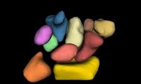
Computational Analysis for Evaluating Joint Morphology and Biomechanics
Our research group is investigating techniques to quantify morphometric and motional characteristics of musculoskeletal tissues, employing in vivo imaging. Our goals are two fold: (1) to allow a comparison of these characteristics across populations based on sex, age and other factors, in the context of disease, and (2) to allow the measurement of changes in tissue shape in conditions where such changes may occur because of the disease process or due to intervention. Applications include wrist instability, osteoarthritis, and neurodegenerative disorders.
References: (1) Foster, A. A. Joshi, M. Borgese, Y. Abdelhafez, R. D. Boutin, and A. J. Chaudhari, WRIST – A WRist Image Segmentation Toolkit for Carpal Bone Delineation from MRI, Computerized Medical Imaging and Graphics; 63:31-40 (2018); (2) B. Foster, C. B. Shaw, R. D. Boutin, C. O. Bayne, R. M. Szabo, A. A. Joshi, and A. J. Chaudhari, A Principal Component Analysis-based Framework for Statistical Modeling of Bone Displacement During Wrist Maneuvers, Journal of Biomechanics, 85, 173-181 (2019)
Molecular Imaging for Skeletal Muscle
Our research group is utilizing molecular imaging methods for the in vivo characterization of glucose metabolism in skeletal muscle. Given the major role skeletal muscle quality plays in a range of disease conditions, such as cancer, musculoskeletal disease, cardiovascular disease, and diabetes, the overall goal of this work is to develop imaging-based biomarkers that can associate with muscle quality and help improve treatment selection and optimization in those conditions.
Reference: C. Zhou, B. Foster, R. Hagge, C. Foster, L. Lenchik, A. J. Chaudhari*, and R. D. Boutin*, Opportunistic body composition evaluation in patients with esophageal adenocarcinoma: association of survival with 18F-FDG PET/CT muscle metrics. Annals of Nuclear Medicine, Dec 2019. (*co-senior authors)
Molecular Imaging for Rapid Screening of Autoimmune Arthritis Therapy
Our research group is utilizing molecular imaging methods, specifically PET, for evaluating the efficacy of autoimmune arthritis therapy in preclinical animal models. The overall goal of this work is to provide means to rapidly screen treatments to identify those that may be effective.
References: (1) A. Mitra, S. Kundu-Raychaudhuri, C. Abria, A. Rona, A. J. Chaudhari, S. P. Raychaudhuri, In vivo quantitative assessment of the therapeutic response in a mouse model of collagen induced arthritis using 18F-fluorodeoxyglucose positron emission tomography, Clinical and Experimental Immunology, 188(2): 293-298 (2017). (2) S. Raychaudhuri, C. Abria, Z. T. Harmany, C. M. Smith, S. Kundu-Raychaudhuri, S. P. Raychaudhuri, and A. J. Chaudhari, Quantitative tracking of inflammatory activity at the peak and trough plasma levels of tofacitinib, a Janus kinase inhibitor, via in vivo 18 F-FDG PET. International Journal of Rheumatic Diseases, 22(12):2165-2169 (2019)